
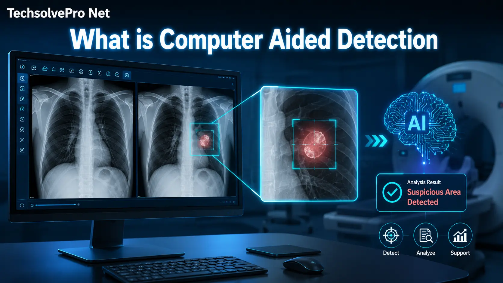
Radiologists review hundreds of images every week and even the most experienced eyes has miss a subtle abnormality buried in a scan. That has exactly the gap this technology was built to close. So what is computer aided detection and why has it become such a trusted second layer of review in hospitals across the country?
At its core this technology uses software to flag suspicious areas on medical images so a physician can take a closer look before finalizing a diagnosis. It doesn’t replace the doctor. It simply catches what tired eyes or a busy schedule might overlook.
What Is Computer Aided Detection?
Computer aided detection often shortened to CADe is a software system. That scans medical images and highlights regions that may need further review. It works alongside mammograms, chest CT scans and chest radiographs to help physicians spot potential disease earlier.
The system haven’t diagnose anything on its own. Instead it marks areas of concern and the radiologist decides whether those marks deserve a second look. This distinction matters because it keeps the physician in control of the final call.
Hospitals started adopting this technology decades ago and primarily in breast cancer screening. Where missing an early tumor can have serious consequences. Since then its use has expanded into lung cancer screening and other imaging applications.
How Does the Technology Actually Work?
The process starts the same way every time. A radiologist reviews the image first using their own training and judgment to look for anything unusual.
Only after that initial review does the software activate. It analyzes the same image using pattern recognition algorithms trained to spot specific features like microcalcifications in breast tissue or rounded nodules in lung scans. Modern AI systems use similar pattern-recognition techniques in many industries. If you’re interested in how intelligent systems learn from data, read our guide on Playing Touhou with Deep Reinforcement Learning.
Once the software finishes its scan then it displays markers on the areas it flagged. The radiologist then goes back through those specific spots to confirm. Whether they warrant further testing or can be dismissed as normal variation.
Why the Two-Step Review Process Matters
This sequence isn’t arbitrary. Requiring the radiologist to review the image before seeing any software markers prevents a kind of tunnel vision where the physician only checks the flagged spots and skips everything else.
Skipping straight to the computer’s markers would risk missing findings the software wasn’t designed to catch. No algorithm identifies every possible abnormality, so the human review always comes first and remains the final word.
Where This Technology Gets Used Today
This detection technology shows up across several major areas of diagnostic imaging, each with its own specific focus and clinical goal.
- Breast cancer screening through digital and film mammography
- Lung cancer screening using chest CT and chest radiographs
- Colon cancer screening through CT colonography for polyp detection
- Cardiovascular imaging for coronary artery disease markers
- Bone metastases detection in whole body nuclear medicine scans
Each application relies on algorithms trained specifically for that anatomy and disease type. A system built for mammography won’t perform the same job on a lung scan, since the visual patterns it searches for are completely different. Anyone researching what is computer aided detection in a specific clinical area should check which anatomy the software was actually trained and validated on.
Computer Aided Detection vs Computer Aided Diagnosis
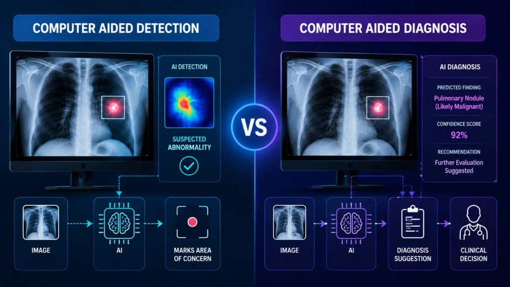
People frequently confuse these two terms but they serve different purpose in a clinical setting. Understanding the difference help clarify, what each system can and cannot do.
| Aspect | Computer Aided Detection | Computer Aided Diagnosis |
| Main goal | Locate suspicious areas | Interpret what a finding likely represents |
| Output | Marks or highlights on an image | Classification, such as benign or malignant |
| Clinical question | Where might something be wrong? | What is this finding likely to be? |
| Regulatory status | Widely FDA approved | More limited approval, still developing |
| Physician role | Reviews flagged areas | Uses classification as additional input |
Detection systems answer the question of where to look. Diagnosis systems attempt to answer what that finding actually means, which is a much harder problem and one still under active development.
How Performance Gets Measured
Not every detection system performs the same and hospitals rely on a few key metrics before adopting one into daily practice.
Sensitivity measures how often the system correctly marks a true abnormality. Specificity often tracked as the number of false marks per image, measures how well the system avoids flagging normal tissue as suspicious.
A system with high sensitivity but poor specificity creates extra work, since radiologists have to sift through many false alarms to find the real findings. The best systems strike a balance between catching real disease and minimizing unnecessary flags.
Benefits Physicians Report in Daily Practice
Clinical studies consistently point to a handful of practical advantages that make this technology worth the investment for many imaging centers.
Radiologists using CADe assistance in breast cancer screening have shown improved detection rates compared to unassisted reads. That improvement have translate into catching cancers at an earlier more treatable stage.
The technology also acts as a safety net during high volume shifts. When a radiologist reviews dozens of studies in a single day. Fatigue naturally increases the chance of an overlooked finding and a second layer of review helps offset that risk.
Screening programs that operate at large scale such as national mammography initiatives. Benefit the most from this consistency. A single missed finding across thousands of screenings can have a real impact on outcomes and even a small improvement in detection rate adds up quickly across a large patient population.
Some imaging centers also report a secondary benefit tied to documentation and quality assurance. Many healthcare organizations now rely on centralized servers to store medical images and patient records securely. Learn more in our guide on What Is Server-Based Computing. Having a recorded set of flagged regions gives departments a way to review edge cases later, which supports ongoing training for newer radiologists learning to interpret difficult images.
Limitations Worth Understanding
No detection system catches everything and physicians are trained to treat software markers as a supplement rather than a substitute for their own judgment.
False positives remain a persistent challenge. A system that flags too many normal findings can slow down workflow and create unnecessary follow up testing for patients who don’t actually need it.
There’s also a real risk in over relying on the technology. If a radiologist starts skipping their own independent review and jumps straight to the flagged areas, genuine findings the software missed could slip through unnoticed.
Cost and integration present another practical hurdle for smaller practices. Adding this software to an existing imaging workflow often requires updated hardware, staff training and ongoing maintenance. Which can be a harder sell for clinics operating on tighter budgets than large hospital systems.
Different patient populations can also affect performance in ways that haven’t always obvious upfront. A system validated primarily on one demographic group may not generalize as well to another. Which is why ongoing evaluation after adoption matters just as much as the initial validation studies.
What This Means for Patients
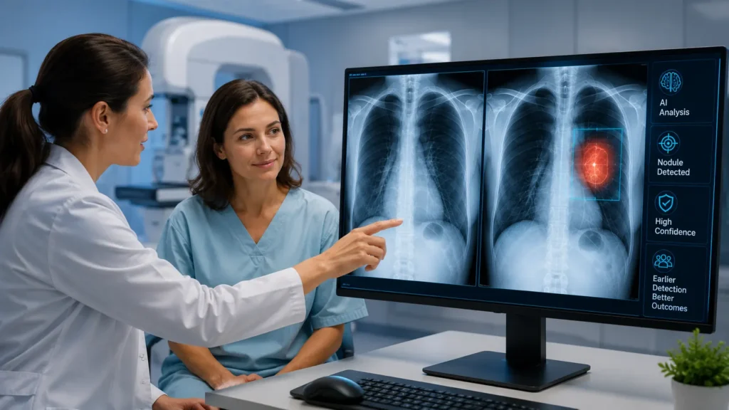
Patients undergoing mammograms, lung cancer screening or colon cancer screening may benefit from this technology without ever realizing it has part of their exam. Medical imaging professionals often spend long hours reviewing scans on computer screens. If you experience facial discomfort during extended computer use, our guide on Nose Pain When Working on Laptop explains common causes and practical solutions. The radiologist still reads every image personally and the software simply adds an extra layer of scrutiny behind the scenes.
This shouldn’t change how patients prepare for a scan or interpret their results. A flagged area doesn’t automatically mean cancer and a clean read doesn’t guarantee nothing was missed. The physician’s final interpretation remains the piece that matters most.
Final Thoughts
Understanding what is computer aided detection comes down to one simple idea: it’s a second set of digital eyes that helps physicians catch what might otherwise slip through. It doesn’t replace clinical judgment and it isn’t perfect but decades of use in breast and lung cancer screening show real value when paired with an experienced radiologist’s review.
FAQs
What does computer aided detection mean?
Computer aided detection means using software to scan medical image and flag area that may need closer review. It helps radiologists catch abnormality they might otherwise miss. Without making any diagnosis on its own.
What is CAD with an example?
CAD or computer aided detection has commonly used in mammography to spot microcalcification and masse that could indicate breast cancer. The software highlights these areas and the radiologist decides whether they need further testing.
What does computer-aided mean?
Computer aided means a computer system supports a task without fully automating it. In medical imaging this means software assists a physician review. Rather than replacing their judgment or making the final decision.
What is an example of a computer aided diagnosis?
An example has software that analyzes a lung nodule on a CT scan and estimates. Whether it looks benign or malignant. Unlike detection which only locates findings. Diagnosis systems interpret what a specific finding likely represents.